Human Vision
Below is a diagram of the eye. Know the labeled parts and what they
do: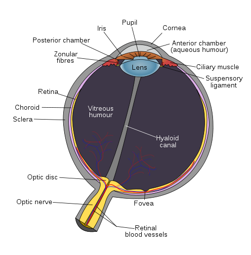
(image: Wikimedia Commons public domain)
Light enters the eye through the cornea, the clear window
at the front. If you wear contact lenses,
they sit
on the cornea. The cornea is made of cells, but is transparent because it has
no blood vessels: the cells get oxygen and excrete waste through tear fluid
bathing them.
Behind the cornea but in front of the lens is the aqueous humor, a transparent, watery fluid, and the iris, the part of your eye that has color (brown, blue or green, usually). The iris is a muscle with a hole in the middle, the pupil. In very bright light, the pupil shrinks, letting less light into the eye (see video below.) In low light, the pupil expands, letting in more light. We say the eye has dark-adapted. That’s why when you are inside a building and then step outside on a bright, sunny day, you are initially dazzled by the brightness.
The lens, behind the iris, serves as the fine-focus for the eye. Unlike camera lenses, this lens is flexible: it can change its shape, and therefore change how much it bends light. However, most of the refraction of the light is done by the cornea, not the lens. That's because it's the air/cornea interface that has a greater difference in wave speed (or said another way, greater difference in index of refraction) than the aqueous humor/lens interface. By the way, this is also why you cannot see well if you open your eyes underwater. When there is water right up against the cornea, there is very little change of n as the light enters the eye, and therefore the light rays are bent hardly at all. Wearing goggles or a mask allows your eyes to work, by having air in front of the cornea like normal.
The eye, when relaxed, is normally focused at infinity. It’s somewhat counterintuitive, but to focus at something close, your ciliary muscles contract, and this releases the tension on the suspensory ligaments attached to the edges of the lens, allowing it to assume its natural curved shape. When you need to focus on something very far away, the ciliary muscles relax, the suspensory ligaments tighten and pull the lens into a flatter, longer-focal-length shape. This automatic process keeps the image sharply in focus on the retina, and is called accommodation.
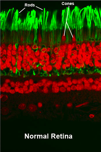
The interior of the eyeball is filled with transparent goo called vitreous humor. The pressure of this stuff keeps the eye in its spherical shape. The back of the eye is covered with the retina, which is the "projection screen" of the eye. The retina is made of zillions of light-sensitive nerve cells called rods and cones. Light hitting the cell causes a shape change in a molecule called rhodopsin, and this change triggers a electrical pulses along the synapse. The machinery of the cell constantly replenishes the rhodopsin so we can continue to see. The cones and rods are excited by the light waves hitting them, which causes them to send electrical pulses along the optic nerve to the brain. They can be stimulated by things other than light, too. For instance, a sharp blow to the head or eye can cause flashes to be perceived. This is why you might "see stars" if you fall and bump your head.
Cones are used for color vision: there are actually three kinds of
cones — those
sensitive to the long-wavelength end of the spectrum, those sensitive to
the short wavelength end, and those sensitive to the middle wavelengths.
These are usually called R, G, and B (for red, green and blue) cones,
respectively, but the ranges of their sensitivity overlap (see graph
below) — the so-called B cones also respond, although not
as strongly, to cyan and green light.
On the other hand, rods are sensitive to all the visible colors, so
they can't distinguish between colors. Their biggest advantage
is that they are more sensitive than cones, so in low light you are using
primarily your rod cells to see. That's why in low light you don't
perceive the colors
of objects very well.

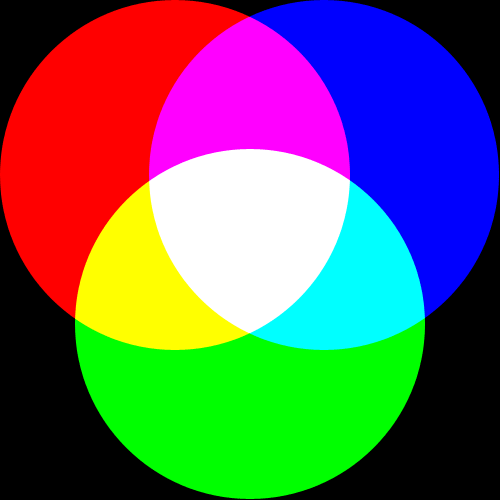
All colors you perceive result from the different response ratios of your R, G, and B cone cells. Take yellow, for example. True yellow light has wavelengths in the neighborhood of 580 nanometers. As you can see from the graph, that wavelength excites both the R and G cones. One way to trick your eye into perceiving “yellow” is to send it a mixture of red and green light. This is exactly what TV screens and computer monitors do. Each pixel of a color CRT or LCD screen is actually a cluster of red, green and blue pixels. Use a strong magnifying glass (a single drop of water placed on the screen works well) to look at the pixels of a TV or monitor. To make a yellow dot, the red and green sub-pixels are turned on, leaving the blue off. This is called additive color mixing, because it involves adding one kind of light to another. Any color can be simulated by some mixture of red, green and blue light, and so these are called the primary colors. This is distinct from the rules of subtractive color mixing you may have learned by mixing paints together in art class, where the pigments in a mixture of paints each absorb (subtract) portions of the light spectrum that is hitting them.
Use this JAVA applet (http://phet.colorado.edu/en/simulation/color-vision) to explore the additive mixing of red, green and blue light to create other colors. Where red and green overlap, the color is yellow; red and blue produce magenta; blue and green produce cyan.
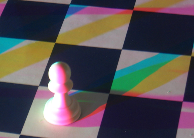
Sometimes, when lighting conditions are right, you may notice odd color shadows. Here's an example. The chess pawn is lit by separate red, green and blue lights, from somewhat different directions. The pawn appears white (on the left side, anyway) because all three primary colors are illuminating it. Notice the shadows. The yellow shadow is where the blue light is blocked, but the red and green lights are NOT blocked. The green shadow is located where the blue light and red light are both blocked.
|
 |
Cones and rods contain light-sensitive chemicals (called rhodopsin in rod cells, and photopsin in cone cells) that absorbs light and trigger electrical nerve impulses, that are ultimately perceived by the brain. When light hits a rod cell, the rhodopsin changes form and is therefore used up. Obviously, the rhodopsin must get replaced by the chemical machinery of the cell. When looking at a bright light, there is a balance between the rhodopsin being used and being replaced, so the cell will have a medium amount of rhodopsin. The cell gets "tired". In darkness, however, the rod cell will have a full load of rhodopsin (because none is being used up) and will respond vigorously when light does fall on it. The cell is "well rested". This process can lead to afterimages: lingering images of high-contrast scenes visible even after the scene has gone away. Perhaps you have dark blotches in your field of vision after accidentally looking into a bright light. Follow the instructions on this PowerPoint presentation to see some interesting afterimages.
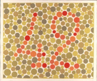
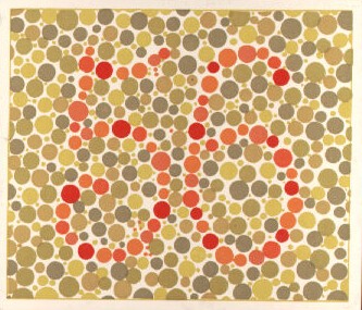
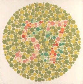
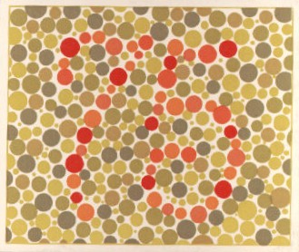
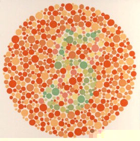
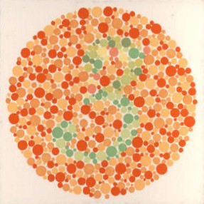
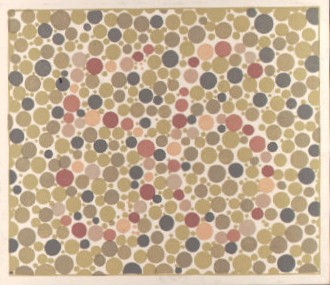
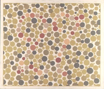
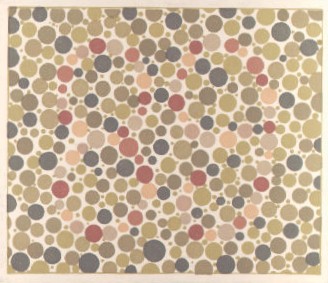
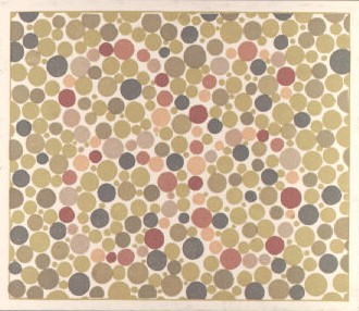
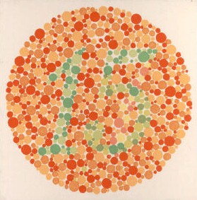
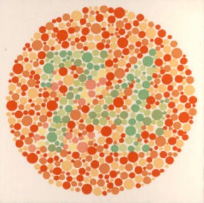
Some people have only two kinds of cone cells, or other differences in the chemistry of the cones, that makes them unable to distinguish some colors, a condition called color blindness. Can you read the numbers in these images? (Click any one of them to see a larger version.)
Over most of the retina, rods outnumber cones 20 to 1. The exception is at the fovea, where there are only cones. The cells are also most closely packed in the fovea, so the part of the image falling on the fovea produces the sharpest picture in your "mind's eye." The fovea is what you use to look at something. The rest of the retina is responsible for your peripheral vision. Because the fovea has no rod cells, when you look directly at something you are using the least light-sensitive part of the retina. If you are trying to see a very faint object in the dark, don't stare. Instead, try to use your peripheral vision: look off to the side. This technique, used by astronomers a lot, is called averted vision.
All the nerves from the retina join together into the optic nerve, and as a bundle are connected to the brain. It’s the optic nerve that keeps your eyeball from rolling away if your eye ever gets knocked out of its socket. The place on the retina where the optic nerve leaves has no cones or rods — it is therefore a blind spot. Normally we don't notice it, but you can "see" your blind spot with this simple test. Close your right eye. With your left eye look straight at the right dot in the box. Move closer or further away. At some distance, the left dot will disappear from your peripheral vision because its image is falling on your retina's blind spot. Once you get it, try again with your other eye.

When you are looking at something and it suddenly disappears, your eyes continue to perceive the image for a short amount of time. In other words, the retina cannot respond instantaneously to changes in the scene it is looking at. This is called persistence of vision. This is imprtant for several technologies. Fluorescent lights flicker 120 times per second. Movies play at 24 frames per second, and standard video plays at 30 frames per second. You don't see any of these flicker because they are just too fast.
Sometimes small bits of tissue break free from the retina and drift about in the vitreous humor. These are called floaters — you can occasionally see them as small out-of-focus specks, especially if you are looking at a bright uniform object like a white wall. Try it. Floaters are perfectly normal — everyone has them.
In some people the eyeball has the wrong shape, or the cornea/lens combination is too weak or too powerful. Either one of these can lead to the in-focus image not being at the proper place, the retina. If the eyeball is too long, then the lens, even in its most stretched state, can't bring the in-focus image to the retina. (Another way of saying this is the cornea/lens are too powerful, converging the light too much.) Only if the object comes very close (and therefore moving the image further from the lens) will the image on the retina be in focus. This is near-sightedness. It can be corrected by wearing a diverging lens in front of the eye. Conversely, if the eyeball is too short, or the refraction by the cornea and lens is weak, the rays won’t converge before they hit the retina, especially if the object is nearby. If this person can see anything clearly at all, it's distant objects: this person is far-sighted. Wearing converging glasses is the solution here, to give the eye a little extra help converging the rays.
Wearing glasses can be a problem when looking through a telescope or microscope: the glasses keep your eye too far from the eyepiece, and reflections off the lenses can be distracting. Near-sighted or far-sighted people can simply remove their glasses and refocus the telescope to compensate. If you have astigmatism (which is kind of like being far-sighted in the vertical axis and near-sighted in the horizontal axis, or vice versa) simply refocusing the telescope/microscope won't work. The solution in this case is contact lenses, or a special eyepiece with long "relief" that allows you to wear glasses and have your eye further back.
In a human eye there are about 120 million rods, and 6 million cones. Compare this to the Wide-field and Planetary Camera on the Hubble Space Telescope, with about 2 million pixels. Clearly, the human eye compares very well with our best technology in terms of number of detectors. In other respects — such as sensitivity, linearity, and spectral response — the human eye is far inferior to the latest electronic detectors. As a result, modern astronomers rarely look through large telescopes, instead relying almost exclusively on CCD chips to do their seeing for them.
It’s amazing that the human eye performs as well as it does, considering
that it is actually poorly designed in several respects. For one thing,
the light-sensitive parts of the cones and rods face the wrong way! The
neural axons (i.e. the ‘wires’ connecting each one, ultimately,
to the optical nerve and the brain) emerge from each cell on the front
side of the retina, so light has to go through them to get to the light-sensitive
parts. The blood vessels feeding the retina are also in front. The eyes
of all vertebrates suffer from these same flaws. The eyes of invertebrates
(such as octopi and squid) are laid out much better: the cells’ light-sensitive
parts face forward, plus the axons are in back and therefore there is
no blind spot. These differences argue strongly that vision evolved independently
in vertebrates and invertebrates.
Activities & Practice
to do as you read
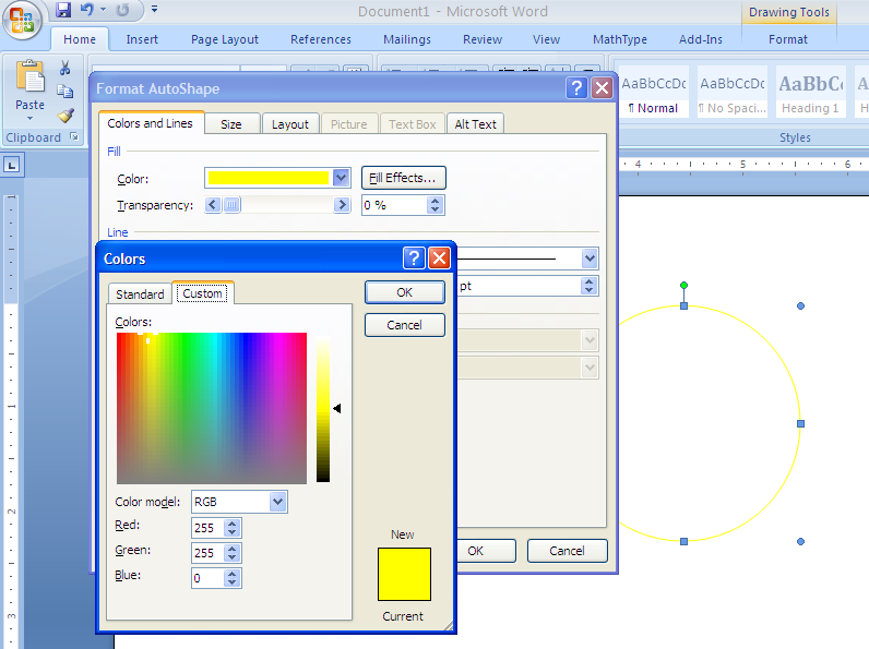
1. Understanding additive color mixing is useful in graphic design. Open a Word document and draw any shape. Then "fill" the shape with a solid color. In the color-selector dialog box, choose "Custom" color. You'll see three numerical input fields labeled R,G, and B, for Red, Green and Blue. Here's a screenshot. Each of these can have any integer value between 0 and 255. Experiment by typing in different values in the input fields, or by simply clicking in the color palette. All serious word processors and graphics programs allow the user this high level of control over color selection.
If you think you might be colorblind, you can read more about it here.
2. Here's a video from NPR's Talk of the Nation: Science Friday, showing how laser eye surgery is used to correct vision.
Here's another video about how the eye and the brain interact.
3. Eye Simulation, including eye defects.
Additional Activities & Practice
1. What kind of glasses lens would you recommend to a patient who is far-sighted, A or B?
2. Take a pair of eyeglasses (either your own or borrowed from somebody
else) and hold them to this page. Is the image upside-down or rightside-up?
Is the
print magnified or reduced? What kind of lenses are they? Is the owner of
the eyeglasses near-sighted or far-sighted?
3. In the book Lord of the Flies, one of the main characters is a near-sighted boy nicknamed Piggy. At one point in the book, a group of boys steals Piggy's glasses and use them to start a fire. What's wrong with this (aside from the fact that stealing is wrong)?
4. Using a ruler and a finger, determine how close you can focus. Put the "zero end" of a ruler against your face, just below one of your eyes, sticking straight out. Close the other eye. Hold up your finger and move it as close as you can, trying to keep it in focus. The closest you can get and still be in focus is called your near point. Repeat the experiment with the other eye and, if you wear glasses or contact lenses, do the experiment with them and without. Is there a significant difference between the nearpoints of your two eyes? If you have glasses or contact lenses, is your nearpoint closer with or without them? By the way, as you age your nearpoint will increase, because the lens of the eye becomes less flexible as the years pass.
5. Close your eyes and face a bright lightbulb. Even though your eyelids are shut, you will still see some light. What color is the light? Why?
6. Do this in a dark area, such as a closet. Take a bright flashlight and shine it in your mouth when your eyes are closed. Make sure no light is hitting your closed eyelids. Surprisingly, you will still see a glow in your field of vision. Verify that the light you are seeing is really coming from the flashlight by turning the flashlight on and off.
(a) Where do you see the light within your field of vision?
(b) Why can you
see any light at all? What color is it, and why?
(c) Why does the light appear
where it does?
7. Take a bright flashlight into the bathroom or another room with a mirror, and put your nose close to the mirror. Turn out the lights, draw the curtains, or do whatever adjustment of the light you need so the room is mostly dark, but there is enough light that you can see one of your own pupils reflected in the mirror. Wait a minute or so for your pupils to dark adapt (grow bigger). Then, turn on the flashlight and reflect its light directly into your eye. Watch how dramatically, and quickly, the pupil shrinks.
8. Here's a very boring movie of the pupil of a human eye dilating over a 15-minute period.